Patent Foramen Ovale: Background and Impact on Divers
Patent Foramen Ovale: Background and Impact on DiversBY DR. NEAL W. POLLOCK
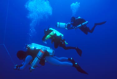
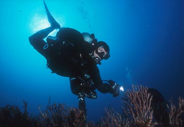
Common anatomical defects of the heart were identified as risk factors for decompression sickness in the 1980s. Between 17% and 35% of the normal population are found to have an anatomical atrial septal defect, or patent foramen ovale (PFO), beyond infancy (1). The incidence among divers who have suffered from serious neurological decompression sickness symptoms was reported to be as high as 61% (2) or 66% (3). This article will present an overview of the condition with emphasis on the implications for the diving population. FETAL CIRCULATIONFetal humans rely on placental circulation and the maternal pulmonary system for gas exchange. Since the growing lungs are not yet functional, they require only enough blood delivery to support their own development. The fetal cardiovascular system has two primary shunts to allow blood to bypass the lungs. It is the interatrial septum that allows blood in the right atrium to flow directly into the left atrium that is the concern of this discussion. Placental circulation is lost at birth and the infant must start breathing independently. After one or more gasps, the previously unused lungs will start to expand. Upon expansion, a redistribution of pressure causes a tissue flap within the left atrium of the heart to be pushed against the septal wall to functionally close the foramen. In the normal course of events, the septal tissues will fuse within the first year after birth to make the closure permanent. However, in a post-mortem review of 965 normal hearts, patent interatrial openings were observed in 27% of the non-infant population (4). EVALUATION OF INTERATRIAL SHUNTS Post-mortem autopsy examination is the most reliable means of identifying anatomical foramen ovale. However, anatomical (or probe) patency does not equate to functional (or physiological or hemodynamic) patency. Being able to work a probe through the cardiac septum at autopsy does not indicate the degree of lateral shunting that was present in the living system. This is why in vivo studies usually report lower incidence rates than post-mortem studies. Functional patency was reported in 9% of 1,000 consecutive patients being scanned by transesophageal echocardiography (5). Functional patency is reliably evaluated using two-dimensional bubble contrast echocardiography (6,7). This technique involves the venous injection of small amounts (5-10 ml) of normal sterile saline solution that has been agitated in the presence of air to ensure the presence of large numbers of microbubbles. Bubbles are highly reflective targets easily detected by the ultrasonic pulses used in echocardiographic instruments. Injections of bubble contrast are made repeatedly during resting breathing while the echocardiograph captures a cross-sectional view of either all four chambers of the heart or the two atria. The microbubbles are clearly visible as they enter the right side of the heart. Functional patency is confirmed if bubbles are seen to cross the septum, and no further testing is conducted. If bubbles are not seen to cross the septum during resting trials, further injections are made during or just prior to the release of the strain phase of a Valsalva maneuver. Injections are repeated until crossover is seen or a pre-established maximum number of test cycles has been reached. As a control, the pressure generated by the Valsalva maneuver will usually be standardized, generally within the range of 40-60 mm Hg above ambient. The dominant pattern of interatrial shunting is left-to-right since the left heart pressure is significantly greater than the right. The Valsalva maneuver is useful in assessing foramen ovale patency by augmenting or prolonging a transient pressure gradient reversal that encourages right-to-left shunting. Septal crossover is usually evident following the release of the strain phase. The presence of a functional patent foramen ovale may have no adverse effect under normal conditions. A minor left-to-right flow would reduce cardiac efficiency, since blood would be sent to the lungs repeatedly, but this may not be problematic. Right-to-left shunting is a greater concern since blood bypasses the lungs and is sent directly through the body (systemic circulation). Minor right-to-left shunts may not affect oxygen content appreciably, but major shunts can limit physical performance. A more insidious problem is the introduction of potentially embolitic materials (e.g., gas bubbles, blood clots) to the systemic circulation that would normally be filtered out in the pulmonary bed. Right-to-left shunting is the suspected agent in a significant number of unexplained or "paradoxical" stroke cases (8). A number of factors may increase right-to-left shunting. These include coughing, the Valsalva maneuver, pulmonary hypertension, chronic obstructive pulmonary disease, and the use of positive pressure ventilation (5). It has also been suggested that a spontaneous reversal of the normal left-to-right pressure gradient may occur during the early phase of ventricular contraction (9). IMPACT OF INTERATRIAL SHUNTS ON THE DIVING POPULATIONDivers face risks from right-to-left shunting beyond those experienced by the non-diving population. The first is a direct result of the action of Boyle's Law. Any bubble present in the systemic circulation is subject to expansion during ascent. Initially non-problematic microbubbles that are shunted systemically could potentially become large enough to cause circulatory blockages. Right-to-left shunting may explain some cases of paradoxical arterial gas embolism (referred to as "undeserved embolism") when classic signs and symptoms are not accompanied by appropriate history or clinical indications of pulmonary barotrauma (10). Interatrial shunting may also alter bubble formation. While the mechanisms of bubble formation and decompression sickness have not been completely resolved, there is strong evidence that micronuclei "seeds" of some nature will initiate or exacerbate bubble formation. Materials shunted right-to-left (that would normally have been filtered out at the pulmonary bed) could serve as "seeds" and increase an individual's susceptibility to bubble formation (2). A substantial bubble load (number and size) may trigger the cascade of events resulting in decompression sickness. The pattern of right-to-left shunting may also be influenced by the activity of diving. It has been demonstrated that the hydrostatic compression of the legs during immersion will increase the cardiac volume prior to contraction (the end diastolic volume or Ôpreload') such that stroke volume may increase up to 30% (11). Right atrial pressure can also be increased by 13 mm Hg (12). The reduced difference between right and left heart pressures may make gradient reversals easier to achieve. Cardiac tissue distension arising from preload increases could also cause tissue distortions that might transiently increase the size and/or patency of the foramen (2). The first major review of foramen ovale patency in divers found that 11 out of 30 divers (37%) treated for decompression sickness had right-to-left interatrial shunts demonstrable by contrast echocardiography. More importantly, the authors reported that 11 out of 18 (61%) who presented with serious signs and symptoms demonstrated shunting during resting respiration (2). An independent group of investigators reported patency in 15 out of 63 divers (24%) in a control group with no history of DCS. This contrasted a patency of 66% (19/29) in divers who had experienced neurological symptoms within 30 minutes of surfacing (3). More recent analyses have employed logistic regression to estimate the relative risk associated with patent foramen ovale. Bove (13) computed a 2.5-fold increase in the odds ratio for developing serious decompression sickness. Schwerzmann et al. (14) suggested a 4.5-fold increase in the odds ratio for developing decompression sickness, but this was a relatively weak study based on retrospective self-reports.One of the issues raised but not resolved in the available literature is the relationship between patent foramen ovale and brain lesions. While it has been suggested that patent foramen ovale is associated with greater numbers of brain lesions (14,15), there is no evidence that this is related to functional deficits.
IMPLICATIONS FOR THE DIVING POPULATION All divers or potential divers should be made aware of the hazards of patent foramen ovale and the availability of testing options. Dive profiles should be selected to minimize bubble formation. Equalizing techniques employing the least Valsalva strain should be used. SUMMARY 1. Lynch J.J., Schuchard G.H., Gross C.M., Wann L.S. "Prevalence of right-to-left atrial shunting in a healthy population: detection by Valsalva maneuver contrast echocardiography." Am J Cardiol 1984; 53(10): 1478-1480. 2. Moon R.E., Camporesi E.M., Kisslo J.A. "Patent foramen ovale and decompression sickness in divers." Lancet 1989; II(8637): 513-514. 3. Wilmshurst P.T., Byrne J.C., Webb-Peploe M.M. "Relation between interatrial shunts and decompression sickness in divers." Lancet 1989; II(8675): 1302-1306. 4. Hagen P.T., Scholz D.G., Edwards W.D. "Incidence and size of patent foramen ovale during the first 10 decades of life: an autopsy study of 965 normal hearts." Mayo Clin Proc 1984; 59(1): 17-20. 5. Fisher D.C., Fisher E.A., Budd J.H., Rosen S.E., Goldman M.E. "The incidence of patent foramen ovale in 1,000 consecutive patients. A contrast transesophageal echocardiography study." Chest 1995; 107(6): 1504-1509. 6. Teague S.M., Sharma M.K. " Detection of paradoxical cerebral echo contrast embolization by transcranial doppler ultrasound." Stroke 1991; 22(6): 740-745. 7. Tsai L.M., Chen J.H. "Abnormal hemodynamic response to Valsalva maneuver in patients with atrial septal defect evaluated by doppler echocardiography." Chest 1990; 98(5): 1175-1178. 8. Hausmann D., Mugge A., Daniel W.G. "Identification of patent foramen ovale permitting paradoxic embolism." J Am Coll Cardiol 1995; 26(4): 1030-1038. 9. Lechat P.H., Mas J.L., Lascault G., Loron P.H., Theard M., Klimczac M., Drobinski G., Thomas D., Grosgogeat Y. "Prevalence of patent foramen ovale in patients with stroke." New Engl J Med 1988; 318(18): 1148-1152. 10. Wilmshurst P.T., Ellis B.G., Jenkins B.S. "Paradoxical gas embolism in a scuba diver with an atrial septal defect." Brit Med J 1986; 293(6557): 1277. 11. Lin Y.C. "Circulatory functions during immersion and breath-hold dives in humans." Undersea Biomed Res 1984; 11: 123-138. 12. Arborelius M. Jr., Balldin U.I., Lilja B., Lundgren C.E.G. "Hemodynamic changes in man during immersion with the head above water." Aerosp Med 1972; 43(6): 592-598. 13. Bove A.A. "Risk of decompression sickness with patent foramen ovale." Undersea Hyperbar Med 1998; 25(3): 175-178. 14. Schwerzmann M., Seller C., Lipp E., Guzman R., Lovbald K.O., Kraus M., Kucher N. "Relation between directly detected patent foramen ovale and ischemic brain lesions in sport divers." Annals Internal Med 2001; 134(1): 21-24. 15. Knauth M., Ries S., Pohimann S., Kerby T., Forsting M., Daffertshofer M., Hennerici M., Sartor K. "Cohort study of multiple brain lesions in sport divers: role of a patent foramen ovale." Brit Med J 1997; 314(7082): 701-705. 16. Cross S.J., Thomson L.F,. Lee H.S., Shields T.G., Jennings K.P. "Safety of contrast echocardiography in screening divers (letter)." Lancet 1990; 336(8730): 1595-1596. 17. James P.B. "Safety of contrast echocardiography in screening divers (letter)." Lancet 1990; 336(8727): 1389-1390.
Copyright ©2004 Global
Underwater Explorers.
All rights reserved. |